Macrophages – the ‘big eaters’ within the immune system – could offer new insights into cell migration if it were possible to accurately visualize their interactions. But researchers still struggle to distinguish overlapping cells in microscope images, which makes it difficult to locate and track these cells. While various methods have been used to segment overlapping cells, a UK team of biomedical engineers, computer scientists and biologists have now worked together to develop an automatic algorithm that could improve these techniques, and also enable more accurate cell location and tracking (MIUA 2017).
The accurate segmentation of biomedical images is a key focus for researchers who are trying to improve image analysis of overlapping cells. José Alonso Solís-Lemus and co-workers at City University of London and King’s College London have now developed an algorithm that improves the detection and segmentation of fluoresced overlapping cells of fruit fly embryos (Drosophila melanogaster). The algorithm, called Anglegram, offers new insights into how macrophages use signals to help them migrate.
In the fluorescence images analysed by the team, the red signal corresponds to the cell nuclei, while the green represents the intracellular proteins, known as microtubules. The key challenge when analysing these images is that the green fluorescence from the microtubules are not strong enough to accurately determine the cell segmentation.
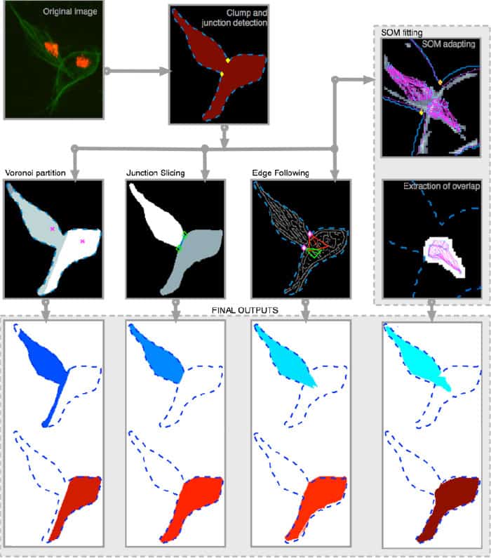
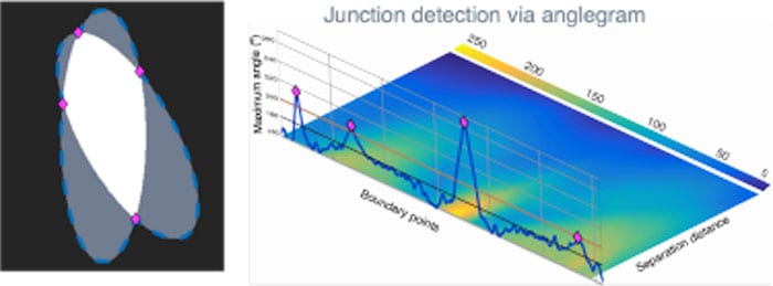
The Anglegram algorithm can help when a cluster of two or more overlapped macrophages is detected by the presence of two or more red nuclei. The algorithm works by automatically detecting the junctions between overlapping cells, as determined by the inner angles of the cell boundaries. This produces a 2D matrix resembling a heat map, which is known as the Anglegram. Boundary points representing the junctions can then be located based on the maximum intensity projection of the Anglegram matrix.
The researchers tested the performance of the Anglegram algorithm by combining it with three different segmentation methods, and comparing the results with a fourth technique that was used on its own as a benchmark. All segmentation methods differ in the way they operate, but the junction slicing technique produced the most accurate segmentation when combined with Anglegram.
The results suggest that Anglegram offers a promising method for improving the segmentation of overlapping cells. Further development of the algorithm could eventually make it possible to detect overlapping cells when more than two cells are clumped together.