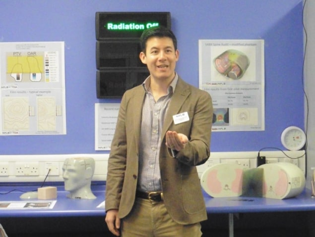
The National Physical Laboratory (NPL), the UK’s National Measurement Institute, has launched the Metrology for Medical Physics Centre (MEMPHYS). The aim of MEMPHYS is to accelerate the development and implementation of innovative diagnostic and therapeutic technologies.
The Centre will provide a range of consultancy and research services, and offer quality assurance and commissioning tools that help get new equipment into routine use, and enable more efficient use of technologies already in the clinic. “MEMPHYS brings together all of the medical physics activities happening at NPL under a single banner and helps us to grow,” explained Rebecca Nutbrown, NPL’s head of metrology for medical physics. “NPL has been expanding its work in this area over the last year.”
One area of medical physics in which NPL already has extensive experience is radiotherapy dosimetry, such as testing the calibration of linacs, for example. “We need to check that the patient treatment is delivered as expected,” said Nutbrown, speaking at the MEMPHYS launch event. “So we devise phantoms that mimic the properties of the human body, embed dosimeters, and then treat them as we would a patient and check the radiation is delivered to the right place.”
For example, as hypofractionated radiation treatments become increasingly prevalent, NPL – in collaboration with the Royal Surrey County Hospital and the Radiotherapy Trials QA (RTTQA) group – used a specialized CIRS head phantom (STEEV) to ensure that all centres using stereotactic radiosurgery (SRS) are treating patients in the same way, and treating accurately. Another development is a dosimetry audit for testing spinal stereotactic body radiotherapy (SBRT) – a treatment that necessitates exceptionally precise beam targeting.
NPL has also created a graphite calorimeter that measures radiation dose extremely accurately. The calorimeter is used to perform measurements on one of NPL’s two linacs; then a hospital will send their instrument to NPL for comparison, thereby ensuring the accuracy of the instruments used for clinical dosimetry.
More recently, NPL has been working on a small portable calorimeter for reference dosimetry of proton therapy. The aim here is to standardize treatments across the UK’s forthcoming proton therapy centres, and to ensure that proton therapy is delivered with the same level of accuracy as conventional radiotherapy.
Lab launch
MEMPHYS also sees the introduction of a new nuclear medicine laboratory. Last December, NPL installed a unique clinical SPECT/PET/CT camera that is directly calibrated against primary standards of radioactivity. As nuclear medicine continues to transition from qualitative to quantitative imaging, the goal is to provide primary standards for emerging quantitative approaches.

The camera will be used to image phantoms, which will then be taken to a clinic for imaging on its scanners. The two data sets, both of which will be processed by NPL to provide quantified measurement uncertainties, are then compared to assess scanner accuracy. To help tailor this process for specific applications, MEMPHYS has also set up a new Rapid Phantom Prototyping Laboratory to produce and optimize medical phantoms.
The lab applies 3D printing to rapidly develop complex phantoms. Examples include anthropomorphic phantoms for medical imaging and dosimetry, and mouse phantoms for calibrating equipment employed in small-animal and radiobiology experiments. The phantoms are fillable with radioactive solutions, though future approaches will involve integrating radioactivity directly into the printing material.

Device development
Finally, NPL’s ultrasound group has developed a new scanner for breast imaging. Ultrasound can be employed as an adjunct to X-ray mammography, and generates more reliable images when breast tissue is dense. The team is developing a novel phase-insensitive ultrasound sensor, originally conceived for a measurement application, to create a system with potentially improved image quality and far fewer artefacts than a conventional ultrasound scanner.
The team has now built a prototype of the phase-insensitive ultrasound CT (piUCT) device. The system employs 14 parallel ultrasound emitter elements, plus one large sensor, all housed in a water tank. The elements can be can moved vertically to select the scan plane. During scanning, the patient lies prone and the breast is imaged without requiring compression, making the procedure much more comfortable than mammography.

The system is currently being evaluated at NPL using a breast phantom, and images will be compared in terms of quality and spatial resolution with X-ray CT and MRI scans. The next step is to test the system on a number of volunteers. For this, the piUCT will shortly be moved to Southmead Hospital in Bristol, for trials on 20-30 patients. Looking further ahead, NPL hopes to partner with a commercial manufacturer to translate this novel system into a clinical device.
Ultimately, MEMPHYS will function as an international centre for excellence, fostering interdisciplinary and inter-sector research to inspire cutting-edge innovations. MEMPHYS will work closely with the NHS, academia and industry to enable the rapid and widespread implementation of a host of new diagnostic and therapeutic technologies.


