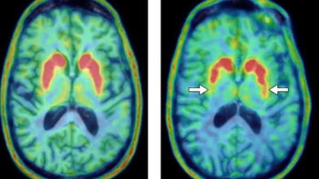
Awake small-animal PET provides a powerful tool to gain unique insight into mechanisms underlying cognition and behaviour. Various methods have been developed that allow brain function imaging on conscious and freely moving rodents, with motion tracking and motion compensation algorithms used to realign the measured lines-of-response.
Uncertainty introduced by noise and jitter, however, and brief periods of fast animal motion with insufficient sampling rate, can reduce the resolution of the motion-corrected images compared with their stationary counterparts. An Australian research team has now devised a method to experimentally measure this residual blurring and use the estimated motion-dependent blurring kernel to improve contrast in the motion-corrected images (Biomed. Phys. Eng. Express 4 035032).
The researchers characterized residual blurring by measuring the point spread function (PSF) in image space using a point source attached to the animal’s optical tracking marker. They hypothesized that the reconstructed PSF should contain information about the animal’s motion, which could then be used to construct a blurring kernel for image deconvolution.
“Point sources have been previously used to measure and mitigate resolution degradation due to physical effects such as parallax errors and positron range,” explained first author Georgios Angelis from the University of Sydney. “Since insufficient motion tracking is yet another factor that degrades image resolution, even when employing motion compensation, we expected that a point source would be appropriate to measure the overall reduction in resolution in motion-affected scenarios.”
Moving phantoms
Angelis and colleagues evaluated their proposed approach by using the microPET Focus220 scanner to image moving phantoms. They constructed point sources from 18F-containing molecular sieves embedded in a cube of tissue-equivalent material. The sources were placed on hot-rod phantoms, which were robotically controlled to perform simple lateral movement, star-like motion and realistic rat head motion. For each pattern, they examined three speed settings.
The researchers reconstructed the emission data using an iterative motion-compensation algorithm. They then manually isolated the point source from the image, modelled it as a mixture of two Gaussian distributions, and extracted the motion-dependent PSF. They observed that the shape of the PSF was dependent on the motion pattern, with lateral motion leading to elongated PSFs and star-like motion giving more rounded PSFs. For both, the width broadened as phantom speed increased.
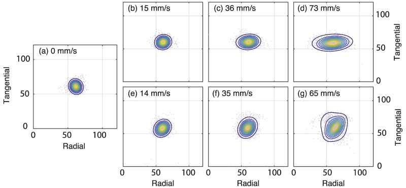
For PET scans with lateral motion at 15 mm/s – a speed typically encountered during an awake rat study – motion-corrected reconstructed images had slightly lower resolution than an image from a stationary acquisition. For moderate (35 mm/s) and fast (70 mm/s) motion, residual motion blurring was clearly visible. Similar findings were seen for the scans with star-like motion.
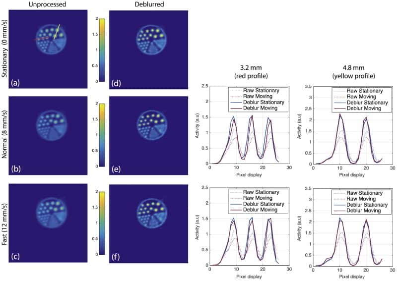
The team then used the PSF within an iterative Lucy-Richardson deconvolution algorithm to mitigate residual blurring in the motion-corrected images. The deblurred hot-rod images appeared highly similar, in terms of resolution and contrast, to their stationary counterparts. In particular, the three larger sets of rods (4.2, 4.0 and 3.2 mm) in the deblurred images appeared more circular compared with the unprocessed images.
To quantify differences in contrast before and after deblurring, the researchers examined line profiles through some of the rods. The profiles showed a clear difference in contrast (peak-to-valley ratio) between the stationary and motion-corrected images. After deblurring, the reconstructed line profiles for stationary and motion-corrected images were highly similar.
Realistic rat motion
The researchers also applied realistic motion (taken from head movements of an awake tube-bound rat) to the phantom using a high-precision robot. Motion parameters included translations and rotations with an average speed of 8 mm/s and maximum speeds of 60-80 mm/s across all three axes. They also emulated a more agitated animal with an average head speed of 12 mm/s.
As before, the motion-corrected images were slightly degraded compared with the stationary acquisition, with resolution deteriorating more for the fast-moving case. Comparing unprocessed and deblurred images demonstrated the improvements in contrast recovery offered by the post processing.
The authors note that although they didn’t show results from a moving animal, it should be fairly straightforward to transfer this technique to their routine freely moving animal experiments. “We have previously performed pilot experiments, where the point source was attached to the optical marker glued on the animal’s forehead, which was well tolerated by the animal,” Angelis noted.
In future work, they plan to incorporate the proposed methodology during image reconstruction (rather than post-reconstruction) using a nested deconvolution approach, which should further improve image quality, primarily due to noise reduction.
“Noise reduction is particularly important for other approaches we are working on, such as direct 4D reconstruction algorithms and deep learning neural networks to derive reliable physiological parameters from the brains of conscious animals,” Angelis explained. “Our overall aim is to improve image quality and parameter estimation for awake and freely moving animal scans, and this approach may provide a straightforward means to overcome basic hardware limitations.”