 Read article: Grounds for celebration as ‘hub of all things coffee’ opens at University of California, Davis
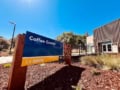
Grounds for celebration as ‘hub of all things coffee’ opens at University of California, Davis
The Coffee Center will be used by more than 50 researchers and includes labs dedicated to brewing, “sensory and cupping” and the chemical analysis of coffee