Researchers from France have combined traditional PET-CT and ultrafast ultrasound imaging (UUI) to create a new hybrid imaging modality that can identify metabolic activities while capturing rapid phenomena with high resolution. This approach should yield simultaneous anatomical, metabolic and functional information while being relatively low in cost. In a proof-of-concept study, the team details how the technology could benefit the fields of oncology and cardiology (Nature Biomed. Eng. 2 85).
Strength in numbers
The current trend in medical imaging consists of coupling imaging modalities to increase the number of parameters obtained during scans. This has led notably to the emergence of PET-CT and SPECT-CT imaging, or more recently to the development of PET-MRI scanners. The advantages of such hybrid modalities, however, are counteracted by their additional cost and added complexities in image processing.
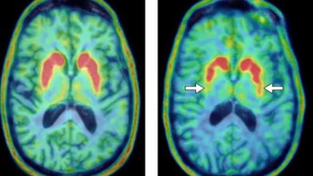
PET imaging allows observation of the metabolisms of radio-tagged molecules such as fludeoxyglucose (FDG), and its combination with CT has long been a reference for cancer imaging as the combination provides both metabolic and anatomical information. However, PET-CT cannot detect dynamic phenomena and is hence only used as a static imaging modality. Conversely, UUI has the unique ability to capture thousands of images per second of 3D volumes, and the development of super-resolution transducers enables the capture of microscopic details such as tissue microvasculature. Combining the three modalities would hence offer unique anatomical, molecular and functional insights.
Merging PET-CT and UUI
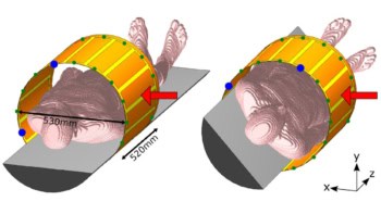
A team from Institut Langevin, INSERM and Université Paris Descartes, led by Bertrand Tavitian and Mickael Tanter, tackled the challenge of designing the first prototype. Their initial work consisted in coupling the UUI transducer probe with the PET-CT scanner. The transducer is positioned over the organ to be imaged and the ensemble is inserted inside the scanner gentry.
Preliminary results showed that the presence of the transducer had negligible impact on the quality of the PET-CT scan, while its location within the gantry-volume yielded a markerless co-registration of the body-of-interest. The next step was to resample the CT, PET and UUI volumes to the same voxel size and fuse them together to obtain the desired representation of the organ.
Providing new insights
The researchers then tested the functionalities of this new modality. For example, they implanted tumour tissues in mice and focussed on the animals’ in vivo metabolism and vascularization, two major hallmarks of cancer used for tumour stratification. The PET-CT scan captured the uptake of the contrast agent FDG by the tumour, and the ultrasound imaging tracked the vessel perfusion. While the analysis revealed an increase in both tumour volume and perfused vessel region over time, their pace of growth differed: the ratio of perfused vessel volume over tumour volume decreased continuously, from 51% on day 14 to 28% on day 32 after implantation.
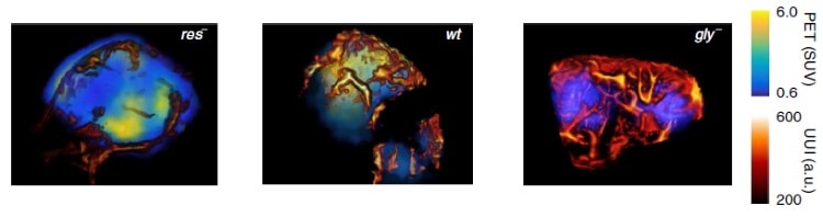
The hybrid imaging modality also facilitated the identification of tumour status based on metabolic and vascular profiles. Indeed, tumours can sometimes affect cells by deregulating their energy metabolism – a feature that can be detected by tracking FDG uptake – while in earlier stages, the metabolism is not altered yet. This difference can guide the choice of treatment given to patients.
The experiment showed that the proposed modality could unambiguously differentiate three different metabolic statuses often found in tumours: an exclusively anaerobic metabolism (res–), and exclusively aerobic metabolism (gly–), and a mixed anaerobic and aerobic metabolism (wt).
Finally, UUI can observe phenomena, such as beating hearts, more easily, and the addition of PET maps of FDG uptake makes delineation of the cardiac anatomy even more precise.
Weighting on the benefits
Combining imaging modalities usually comes with extra development complexity and cost. In this respect, the integration of a relatively inexpensive and portable ultrasound transducer probe in a PET-CT scanner is appealing. The fact that PET-UUI is simultaneous allows co-registration of the volume imaged and the establishment of a topographical relationship between multiple imaging parameters.
It is too early to know whether such an imaging combination will power new breakthroughs, but its potential for linking physiology and pathophysiology is promising. It could become a new standard for cancer imaging, where its ability to characterize both metabolic and vasculature can be particularly helpful.